![]()


Our group specializes in morphological, molecular, and experimental pathological studies of liver tumors, especially primary liver cancer. Our recent research themes include (1) the relationship between cancer stem cells and carcinogenesis or progression of liver cancer, (2) the relationship between chronic hepatic damage and carcinogenesis mechanism, (3) identification of molecules related with portal vein invasion of hepatocellular carcinoma (HCC), (4) histogenesis of combined hepatocellular cholangiocarcinoma (CHC), (5) identification of histological diagnostic marker of CHC, (6) studies of gene expression and genetic abnormalities of CHCs, especially cholaniolocarcinoma, and (7) basic studies of molecular targeted therapy of HCC with new drug delivery system and/or new drugs.
To perform these studies, we collected tissue samples from several hundred surgically-resected liver cancer cases over 20 years. Tissues of cancerous and non-cancerous areas were processed and prepared into FFPE samples and frozen samples, which are stocked with macroscopic pictures and clinical information. We also have originally-established 15 hepatobiliary cancer cell lines (e.g., 11 HCC cell lines, 2 CHC cell lines, and 2 cholangiocarcinoma cell lines), one extrahepatic bile duct carcinoma cell line, and one gallbladder carcinoma cell line in our institute. HCC cell lines include two clonally-related, morphologically and biologically distinct human HCC cell lines established from a single nodule, which is useful to study the mechanisms of dedifferentiation and progression of HCC. In addition, three HCC cell lines were established from peritoneal effusion of patients in the terminal stage. We have established and maintained two rare CHC cell lines. One of these CHC cell lines was established from a CHC of typical type and the other was established from a CHC, subtype with stem cell features, intermediate-cell type.
We not only perform clinicopathological studies of liver cancer based on the detailed observation of HE-stained sections and clinical data, but also molecular pathological studies based on immunohistochemically-stained sections showing expressions of various kinds of molecules. We also perform cDNA microarray analysis of RNAs extracted from the areas of interest in FFPE or frozen sections with macro- or micro-dissection method, compare gene expression, and identify potential key molecules. Then, we perform immunohistochemical analysis of potential key molecules to investigate their significance. If necessary, liver cancer cell lines are used to examine molecular function. We employ this procedure to identify the molecules related to the pathological diagnosis of rare primary liver cancers (e.g., CHC) and those related to characteristic biological behaviors (e.g., portal vein invasion) of HCC and other tumors.
In conclusion, we apply pathological approaches to contribute to the identification of histological diagnostic and biological markers, as well as the development of effective therapeutic strategies in liver cancer.
Hirohisa Yano, M.D.,Ph.D.


- Usefulness of Tumor Tissue Biopsy for Predicting the Biological Behavior of Hepatocellular Carcinoma
-
Taro Shioga, Reiichiro Kondo, Sachiko Ogasawara, Jun Akiba, Shinji Mizuochi,
Hironori Kusano, Yutaro Mihara, Masahiko Tanigawa, Yoshinao Kinjyo,
Yoshiki Naito, Ryoko Kuromatsu, Osamu Nakashima and Hirohisa YanoBackground/Aim: Assessment of the biological behavior of tumors is important for choosing an appropriate cancer therapy. In hepatocellular carcinoma (HCC), the biological behavior can be assessed by tumor morphology and molecular biology. This study investigated the usefulness of tumor tissue biopsy for predicting the biological behavior of HCC.
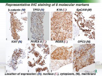
Patients and Methods: We studied 43 patients who underwent hepatectomy and preoperative liver tumor biopsy for HCC. We performed clinicopathological and immunohistochemical (IHC) analyses. The expression of the following molecules was examined: regulator of G-protein signaling 5 (RGS5), glypican-3 (GPC3), keratin 19 (K19), epithelial cell adhesion molecule (EpCAM), protein induced by vitamin K absence or antagonist-II (PIVKA-II), β-Catenin, and p53.
Results: There was an overall 83.7% agreement regarding tumor differentiation between the preoperative biopsy specimens and the resected specimens. The accuracy of IHC analysis was more than 70% for all molecules between the preoperative biopsy specimens and the resected specimens. The RGS5- positive biopsy cases had higher serum α-fetoprotein levels (p=0.04), a higher rate of moderately or poorly differentiated tumors (p=0.02) and portal vein invasion (p=0.0003) than the RGS5-negative biopsy cases. The GPC3-positive biopsy cases were younger (p=0.04), had higher serum PIVKA-II levels (p=0.01), and a higher rate of portal vein invasion (p=0.03) than the GPC3-negative biopsy cases. The PIVKA-II-positive biopsy cases had significantly higher serum PIVKA-II levels than the PIVKA-II-negative biopsy cases (p=0.02). The other molecular markers showed no significantly different clinical findings between the positive and negative cases.
Conclusion: In HCC, there was a high agreement rate of both the histopathological and IHC findings between preoperative biopsy specimens and resected specimens. In the biopsy specimens of HCC, RGS5 and GPC3 expression were useful molecular makers for predicting portal vein invasion. Liver tumor biopsy is useful for predicting the biological behavior of HCC through histopathological and immunohistochemical findings.