![]()


The main aims of our Liver Cancer Research Division are to conquer difficult-to-treat cancers such as liver cancer and pancreatic cancer, and to regenerate cirrhotic liver caused by chronic hepatitis C virus infection. Projects related to cancer treatment include 1) development of tumor tissue-specific anti-angiogenic treatments through regulating tumor endothelial cell-specific microRNAs, 2) establishing precise diagnosis of early pancreatic cancer by analyzing microRNAs derived from exosomes in pancreatic juice, 3) investigating the potential involvement of T-cell factor-4 isoforms in regulating cellular phenotype of cancer stem-cell-like cells, 4) unveiling novel functions of the immune check point molecule PD-L1 in sarcomatous liver cancer, 5) modulation of liver cancer immunity by multi-kinase inhibitors, and 6) studying the usefulness of IGFBP-1 as a biomarker in the treatment of advanced hepatocellular carcinoma with molecular-targeted drugs. Projects related to cirrhotic liver include a clinical trial for regeneration of cirrhotic liver using CD34-positive cells in humans, and an experimental challenge using iPS-derived hepatocyte-like cells and endothelial cells for the same purpose.
In November 2017, Kurume University obtained a 5-year grant as part of the research branding project at private universities supported by the Ministry of Education, Culture, Sports, Science and Technology. Although the branding project finished in March 2020, we have discovered promising findings in research project No. 2 that will go to patent application in the near future.
Hironori Koga, Division Chief


- A. Exploration of T-cell factor-4 isoform-dependent dysregulation of Wnt signaling in liver cancer cells
-
Hironori Koga
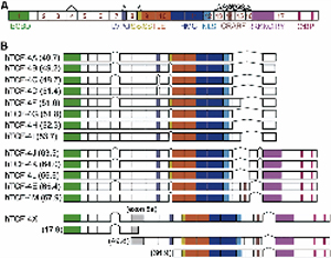
Previously we identified 14 isoforms of T-cell factor (TCF)-4, a key transcriptional factor in the Wnt signaling pathway, from human hepatocellular carcinoma (HCC) cell lines (Figure 1) (Exp Cell Res 2011). Functional analysis of the isoforms demonstrated that the SxxSS motif, that localizes at the head of exon 9, played crucial roles in increased cell proliferation and resistance to hypoxia (PLoS ONE 2012). It also found that loss of exon 4 contributed to an increase in resistance to chemotherapeutic agents and epithelial-mesenchymal transition (Liver Int 2013). These findings suggest that expression levels and variation of TCF-4 isoforms tightly regulate diverse phenotypes of liver cancer cells. However, our methods using classical gel electrophoresis had limitations in quantitatively measuring those expression levels. To overcome these limitations, we have established a novel peptide nucleic acid-based RT-PCR system, and have completed testing the potential of the system in assessing expression level in various isoforms in several cancer cell lines, including liver, stomach, pancreas, and colon cancer cells. Preliminary results are as follows: 1) gastric and colon cancers expressed high levels of TCF-4J, suggesting a possible involvement of the TCF-4J isoform in activation of the Wnt signals in digestive tract cancers. 2) combined hepatocellular-cholangiocellular carcinoma did not express high levels of TCF-4J, although this type of cancer had an adenocarcinoma-like component. Currently, we are identifying unique interactions between TCF-4 isoform profiles and cancer phenotypes.
- B. CD34-positive cell transplantation improves liver fibrosis in a mouse model of non-alcoholic steatohepatitis
-
Toru Nakamura, Atsutaka Masuda et al.
Background: We have already reported that CD34+ cell transplantation was useful for antifibrosis and regenerative therapy in carbon tetrachloride-induced liver cirrhotic mice and against hepatitis virus-related liver cirrhosis in humans. Non-alcoholic steatohepatitis (NASH), characterized by steatosis with inflammation and liver cell injury, can progress to liver cirrhosis. The aim of this study was to investigate the efficacy of cell transplantation therapy with ex vivo-expanded mouse CD34+ cells in a diet-induced NASH model.
Methods: Mouse CD34+ cells were isolated from mouse bone marrow mononuclear cells by magnetic cell sorting and cultured in StemSpan-SFEM medium supplemented with VEGF, SCF, IL-6, Flt-3 ligand and TPO for 7 days. Ex vivo-expanded CD34+ cells were evaluated by FCM analysis. C57BL/6J mice were fed a control (10% kcal fat) or a choline-deficient amino acid-defined, high-fat diet (CDAHFD; 60% kcal fat, 0.1% methionine) for up to 12 weeks. At week 9, the mice were divided into 5 treatment groups: Normal food (NF) + saline-infused (group A); NF + single CD34+ cell transplanted (group B); CDAHFD60 + saline-infused (group C); CDAHFD60 + single CD34+ cell transplanted (group D); CDAHFD60 + repeated CD34+ cell transplanted (group E). We analyzed routine liver function tests and liver fibrosis related mRNA expression, and measured the morphometry of fibrotic areas by Azan-Mallory staining.
Results: After 7-day culture, CD34+ cells were effectively expanded. The expanded CD34+ cells were also characterized as positive for cell surface markers of CD31 and CD34, whereas they were negative for F4/80. Mice on CDAHFD developed liver fibrosis and AST, ALT and T-Bil values were significantly higher in group C than in groups A and B. Repeated expanded CD34+ cell transplantation reduced liver fibrosis. Semiquantitative analysis indicated that the relative extent of the fibrotic area was 5.2 ± 0.3% in group E compared with 7.4 ± 0.6% in group C. Real-time PCR showed that the mRNA expression of Col1a1, Acta2, TGFb1 and Timp1 was significantly higher in group C than in groups A and B; however, repeated expanded CD34+ cell transplantation significantly decreased the mRNA expression of Col1a1 and tended to down-regulate the mRNA expression for Timp1 compared with levels measured in group C. The ratio of the liver/body weight in group E was significantly higher than that in group C (8.6 ± 0.2% vs 7.5 ± 0.2%, respectively).
Conclusion: Repeated expanded CD34+ cell transplantation may have beneficial effects on fibrosis progression in NASH.
Figure. Microphotographs of liver sections.
Fibrosis was less notable in single and repeated expanded CD34+ cell transplantation than in saline-infused livers according to Azan-Mallory staining. The percentage of the fibrotic areas was significantly less in repeated expanded CD34-treated livers (group E) than in saline-infused livers (group C). Bar = 100mm.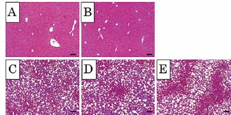
- C.The functional analysis of laminin receptor for hepatic fibrosis and development of hepatocellular carcinoma in non-alcoholic steatohepatitis.
-
Takafumi Yoshida
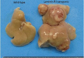
Background and Aims: We have previously reported that high expression of laminin receptor (LR) in hepatocellular carcinoma was associated with an aggressive clinicopathological phenotype including high histological grade and the presence of portal invasion. Furthermore high LR expression in the background liver was an independent prognostic factor and was significantly correlated with lower serum albumin levels. In this study we evaluated whether high expression of LR expression promotes hepatic fibrosis and development of hepatocellular carcinoma in non-alcoholic steatohepatitis. Methods: Mice expressing LR (LR-Tg mice) and control mice were subcutaneously injected with streptozotocin at 2 days of age and 4 weeks later fed with high fat diet leading to steatohepatitis with insulin resistance. Effects of ectopic expression of LR on steatohepatitis were histologically and biochemically evaluated. Results: Immunohistochemistry stains and quantitative real-time PCR revealed that ectopic expression of LR significantly increased hepatic inflammation and fibrosis in accordance with histologically findings. The developments of HCC was also promoted in the livers of LR-Tg mice compared to that of control mice. Conclusions: The present results demonstrated that high expression of LR promotes not only the progression of fibrosis but also the development HCC in steatohepatitis. LR is a potential therapeutic target for steatohepatitis.
- D.Involvement of cancer associated fibroblast in lenvatinib resistance for hepatocellular carcinoma
-
Hideki Iwamoto
Background:
Lenvatinib (Len) has now become a first-line therapy for unresectable hepatocellular carcinoma (HCC). Len inhibits tumor angiogenesis by blocking not only VEGF signaling, but also FGF signaling. Len achieves a high therapeutic response, but HCC gradually acquires Len resistance during the course of treatment. Cancer associated fibroblast (CAF) is an important cell component within the tumor microenvironment that is known to be correlated with drug resistance and tumor progression. In this study, we examined involvement of CAF in acquisition of drug resistance to Len treatment for HCC using hepatoma mouse model.Methods:
We examined changes in the tumor microenvironment in tumors treated with sorafenib and Len using a hepatoma mouse model. Change of genetic expression in CAF was also examined after cell isolation using magnetic beads. We also evaluated direct changes in genetic expression in Len using a fibroblast cell line.Results:
Sorafenib and Len significantly inhibited tumor angiogenesis in this mouse hepatoma model. Moreover, α-smooth muscle actin (SMA) positive area, which revealed a stromal component mainly consisting of CAF, was significantly increased after sorafenib and Len treatment. The expression of hepatocyte growth factor (HGF) and its receptor c-MET was significantly increased after Len treatment (Figure 1). CAF component isolated by magnetic beads showed increased HGF expression level. In the in vitro experiment, addition of Len into the medium resulted in a direct increase of HGF in a fibroblast cell line. Inhibition of FGF signal directly induced HGF expression in the fibroblast cell line.Conclusion:
The present study shed light on the mechanism of drug resistance to Len in HCC. Len induces tumor hypoxia, which resulted in increased c-MET expression in HCC. On the other hand, Len directly induces an increase of HGF in CAF. The study suggested that activation of the HGF-c-MET axis due to Len contributed to development of drug resistance to Len in HCC.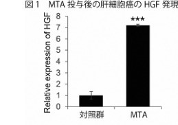
- E.Creation of an innovative DDS formulation for the treatment of NASH
-
Atsutaka Masuda, Toru Nakamura
Background: We have already reported that CD34+ cell transplantation was useful for antifibrosis and regenerative therapy against carbon tetrachloride-induced liver cirrhosis in mice and hepatitis type-C virus-related liver cirrhosis in humans. Those results showed that transplanted CD34+ cells selectively homed and engrafted to the cirrhotic liver. The aim of this study is to create a bio drug delivery system that utilizes the homing ability of CD34+ cells to deliver therapeutic agents for Non-alcoholic steatohepatitis (NASH) to the liver. Therefore, we created an in vivo model that can calculate the liver engraftment rate of transplanted cells, and examined engraftment of CD34+ cells using adipose-derived mesenchymal stem cells (Ad-MSCs) as a control.
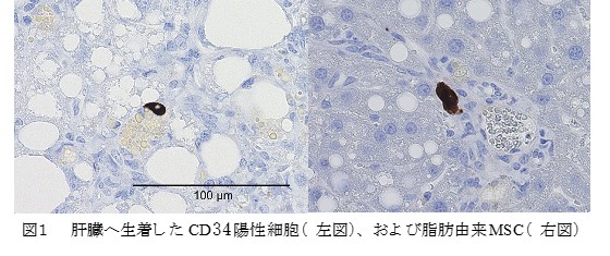
Methods: Mouse CD34+ cells were isolated from GFP-tg mouse bone marrow mononuclear cells using a magnetic cell separation system and cultured in StemSpan-SFEM medium supplemented with VEGF, SCF, IL-6, Flt-3 ligand, and TPO for 7 days. Ad-MSCs were harvested from mouse inguinal subcutaneous fat and cultured in 10% FBS/DMEM F12 medium for 5 passages to purify. Cultured CD34+ cells and Ad-MSCs were evaluated by FCM analysis. Six-week-old male C57BL/6J mice were fed a normal diet or a choline-deficient methionine reduced 60% fat diet (CDAHFD60) for 16 weeks. Both types of cells were transplanted transplenically, and 2 days after transplantation, all mice were slaughtered. Livers were harvested, measured for liver volume, and fixed in formalin. The liver sections were immunostained with anti-GFP antibody, the number of GFP-positive cells per area was counted, and the cell length diameter of GFP-positive cells was measured to calculate the estimated liver engraftment rate of the transplanted cells.
Results: FCM analysis showed that cultured CD34+ cells were CD31+, CD45+, and F4/80-, while Ad-MSCs were CD44+, CD90+, CD45-, and CD31-, and the positivity of CD73 and CD105 was slightly lower than previously reported. The GFP-positive cell length diameters observed in liver sections were significantly different; CD34+ cell: 9.3 μm, Ad-MSC: 33.8 μm. The calculated liver engraftment rate of cultured CD34+ cells was significantly increased in the CDAHFD60 diet group compared with the normal diet group (38.5% vs. 17.5%), and was also significantly higher than that of Ad-MSCs (38.5% vs. 30.2%).
Conclusion: We have developed an in vivo model that can calculate the liver engraftment rate regardless of the cell length diameter. CD34+ cells homed more strongly to the liver exerting by the metabolic stress and this homing ability of CD34+ cells was considered to be stronger than that of Ad-MSC cells.
- F.Establishment of rapid and accurate diagnostic assay with cancer-specific microRNAs derived from exosomes in patients with pancreatic cancer
-
Takahiko Sakaue, Hideki Iwamoto, Hironori Koga
Although pancreatic ductal adenocarcinoma (PDAC) is one of the malignant tumors with extremely poor prognosis, good prognosis can be expected if early detection and treatment are possible. Several studies have reported that exosomes in blood may have potential as novel biomarkers, but there have been no practical applications to date.
The aim of this study was to search and identify tumor-specific exosomal microRNAs from pancreatic juice which directly contact with PDAC in the pancreatic duct, and to diagnose precancerous lesions and early-stage pancreatic cancer, which cannot be recognized by imaging tests, with a sensitivity and specificity superior to those of known tumor markers and pancreatic juice cytology. We succeeded in identifying two pancreatic cancer-specific microRNAs derived from exosomes in pancreatic juice using our original method. Subsequent validation using 15 cases of pancreatic juice from patients with pancreatic cancer and 11 cases of pancreatic juice from patients with chronic pancreatitis showed higher sensitivity, specificity, and positive diagnosis rate than existing tumor markers and pancreatic juice cytology. Furthermore, by adding these two microRNAs to the pancreatic juice cytology, it was shown that the microRNAs are useful as biomarkers for the diagnosis of pancreatic cancer. In addition, a study on the application to serum diagnosis in a small number of cases showed promising results. Furthermore, the fact that the expression of microRNAs in exosomes was decreased in serum after curative resection of pancreatic cancer suggested that the microRNAs may be useful for monitoring the effect of treatment. We are currently conducting a functional analysis of these exosome-derived microRNAs.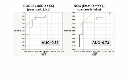
- G.Anti-PD-L1 antibodies promote proliferation via AXL in sarcomatous liver cancer cells
-
Toshimitsu Tanaka, Hironori Koga
Background: Immune checkpoint inhibitors (ICI) are a powerful tool to treat advanced malignant tumors. However, they can sometimes induce so-called hyperprogressive disease (HPD) in patients with such tumors. To reveal the underlying mechanism of HPD, we assessed possible direct action of ICIs on PD-L1-expressing liver cancer cells under immune cell-free condition, focusing on interaction of molecules involved in the proliferation.
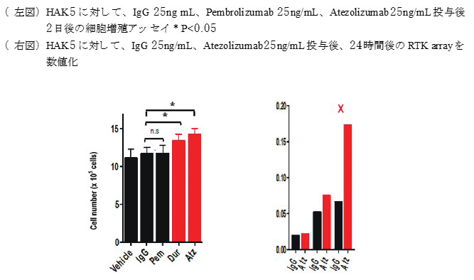
Materials and Methods: Eleven human liver cancer cell lines, including Huh-7, Hep3B, HepG2, HLF, HAK-1A, HAK-1B, KMCH-1, KMCH-2, KYN-1, KYN-2, HAK-5, and one human fetus-derived immortalized hepatocyte cell line (OUMS-29) were used in this study. Expression analysis of protein and mRNA was done by western blotting and real-time PCR, respectively. Gene silencing and overexpression of PD-L1 in HAK-5 and proliferative activity of HepG2 cells after treatment with anti-PD-L1 antibodies (durvalumab (Dur) and atezolizumab (Ate)) was evaluated by cell counts. Molecules involved in the anti-PD-L1 antibody-induced cellular proliferation were evaluated using the phosphorylated RTK Array Kit.
Results: Of 12 cell lines, the sarcomatous liver cancer cell line HAK-5 demonstrated extremely high expression of PD-L1 at both protein and mRNA levels. When HAK-5 cells were treated with anti-PD-L1 antibodies (durvalumab (Dur) and atezolizumab (Ate)) for 48 h, an increase of about 20% in cell proliferation was found compared with those treated with IgG and pembrolizumab, an anti-PD-1 antibody. PD-L1-nonexpressing Huh-7 and HepG2 cells did not respond to Dur or Ate. Gene silencing and overexpression of PD-L1 in HAK-5 and HepG2 cells led to a significant decrease and increase in cell proliferation, respectively. In the RTK array, the treatment with Ate increased expression of X.
Conclusion and Future Direction: Our study found that HLF and HAK5 were expressed in PD-L1 in human liver cancer cell lines and that treatment with anti-PD-L1 antibodies enhances cell proliferation ability. Moreover, it was revealed that higher expression levels of PD-L1 correlated with faster proliferation speed in liver cancer cells. X was identified as a molecule involved in the anti-PD-L1 antibody-induced cancer cell proliferation . In the future, we will elucidate the co-working mechanism of anti-PD-L1 antibodies and X.
- H.Molecular targeted agent alters tumor immune microenvironment from ‘cold’ to ‘hot’ in hepatocellular carcinoma
-
Hiroyuki Suzuki, Hideki Iwamoto
Background
Molecular targeted agents (such as sorafenib and lenvatinib) have become a standard therapy for advanced hepatocellular carcinoma (HCC), however, their effect can still be limited. Recent studies combining bevacizumab, one of the molecular targeted agents, with atezolizumab, one of the immune checkpoint inhibitors (ICIs), have reported promising results for patients with HCC. However, the mechanisms of these combination therapies have not been fully clarified. HCC is known as an immune ‘cold’ tumor, in which immune cell infiltration is low. Several studies demonstrated that immune ‘cold’ tumors were resistant to ICIs, whereas immune ‘hot’ tumors were sensitive to them. Therefore, to alter tumor immune microenvironment from ‘cold’ to ‘hot’ could be an important therapeutic strategy for HCC in combination with ICIs. Here we show that a molecular targeted agent could alter the tumor immune microenvironment from ‘cold’ to ‘hot’.Method
We established an immune syngeneic HCC mouse model by transplanting a hepatoma cell line (derived from C57BL/6) subcutaneously or orthotopically into C57BL/6 mice followed by oral treatment with molecular targeted agent for 4 weeks. We then performed immunohistochemistry to evaluate alteration of tumor immune microenvironment caused by molecular targeted agent.Results
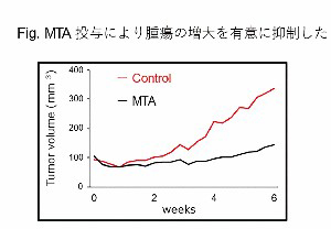
Tumor volume was significantly suppressed in the molecular targeted agent group compared to the vehicle group. Immune cells were scarcely detected in the vehicle treatment group, which suggested that the tumor immune microenvironment of the syngeneic HCC mouse model was an immune ‘cold’ tumor. Immune cells such as CD4 positive and CD8 positive lymphocytes were significantly increased in the molecular targeted agent group as compared to the vehicle group. These results suggested that the molecular targeted agent altered the tumor immune microenvironment from ‘cold’ to ‘hot’.Conclusion
In this syngeneic xenograft mouse model for HCC, a molecular targeted agent apparently altered the tumor immune microenvironment from 'cold' to 'hot'. The findings obtained from this study support the therapeutic concept of combining drugs targeting angiogenesis and immunology against immune ‘cold’ HCC. Further studies will focus on analyzing the lymphocytes which were changed by the molecular targeted agent.