![]()


The Molecular Targeting Division currently consists of 10 groups (Internal Medicine, Gastrointestinal Surgery, Hepatobiliary and Pancreatic Surgery, Obstetrics and Gynecology, Neurosurgery, Orthopedics, Radiology, Hematology and Oncology, Pathology, and Diagnostic Pathology). Each group works independently and conducts its own distinctive research activities. Several groups are actively investigating immune checkpoint inhibitors, which are now considered to be the fourth standard treatment for cancer, and the tumor immune environment. In addition, studies on the proper handling of samples for genome sequencing, and analyses of cancer genome data to identify novel biomarkers are being carried out. These studies reflect actual current medical needs and are expected to produce results that can be applied to clinical treatment. Furthermore, investigations using iPS cells and cultured cells are also being conducted, and are expected to expand from bench to bed in the future.
The goal of the Molecular Targeting Therapeutic Division is to identify synergies among the results obtained by each of our various groups and to find ways to translate these results into cross-organ cancer treatments.
Jun Akiba, MD


- The Diversity and The Shared TCR Repertoire Analysis in Esophageal Squamous Cell Carcinoma
-
The tumor immune response is dependent on the interaction between tumor cells and the T-cell subset expressing the T cell receptor (TCR) repertoire that infiltrates into the tumor microenvironment. Here, we explored the diversity of TCR repertoires, while also looking for shared TCR repertoires expressed on the surface of locoregional T cells. We aimed to identify the T lymphocyte subsets infiltrating into esophageal squamous cell carcinoma (ESCC) in order to predict the efficacy of immunotherapy and hopefully develop a novel immune-oriented therapeutic strategy. Fifty-three patients were enrolled in this study, and immunohistochemical analysis of CD3, CD8, CD45RO, FOXP3, CD274, HLA class I, and AE1/AE3 was performed. Digital pathological assessment was performed to evaluate the expression level of each marker. The clinicopathological significance of the immune-relation high (IR-Hi) group was assessed. Adaptor ligation polymerase chain reaction and next-generation sequencing were performed to explore the diversity of the TCR repertoire and to investigate the shared TCR repertoire in the IR-Hi group. Repertoire dissimilarity index (RDI) analysis was performed to assess the diversity of TCR, and the existence of shared TCRα and TCRβ was also investigated. Further stratification was performed according to the expression of markers of different T cell subsets. Patients were stratified into IR-Hi and immune-relation low (IR-Lo) groups. Cancer-specific survival and recurrence-free survival were significantly better in the IR-Hi group. The diversity of the TCR repertoire was significantly higher in the IR-Hi group. TCR repertoire analysis revealed 27 combinations of TCRα and 23 combinations of TCRβ VJ regions that were shared in the IR-Hi group. Overall, our findings showed the existence of a IR-Hi group and IR-Lo group, and we found that the IR-Hi group maintained the diversity of TCR. It appears possible that a portion of these cases may share TCRαβVJ repertoires. The prognosis of ESCC was affected by the existence of immune response cells and could possibly be stratified by the T cell subsets.
Keywords: Comprehensive T cell receptor repertoire analysis, esophageal squamous cell carcinoma, TCR diversity, shared TCR
- Elucidation of anticancer drug sensitivity / resistance related genes based on genome function
-
Background: When considering cancer treatment, sensitivity and resistance to anticancer agents are important issues. Although a large number of genes involved in anticancer drug sensitivity and resistance are thought to exist, most have yet to be sufficiently elucidated.
Purpose: The purpose of this study will be to elucidate the group of genes involved in anticancer drug sensitivity / resistance by means of a comprehensive search of functionally responsible genes using a large-scale gene trap insertion mutant cell library. Specifically, we will search for and analyze functional groups of genes involved in sensitivity and resistance for two types of cytotoxic anticancer drugs, Oxaliplatin and Irinotecan.
Research Plan and Method: (1) Enrichment and isolation of anticancer drug insensitive mutant cells : Anticancer drug treatment of a large-scale gene trap insertion mutant cell library and amplification of surviving cells will be repeated three times to create a pool of anticancer drug insensitive mutant cells. In this cell pool, it is expected that a large number of mutant cells with reduced anticancer drug sensitivity due to gene trap insertion mutation will be included. Isolation of mutant cells will be carried out by diluting and culturing these cell pools. (2) Elucidation of genes in mutant cells : The expressed genes from 8 clones of anticancer drug mutant cells and 4 clones of wild type cells will be analyzed by 5’-RACE / sequence / BLAST analysis.(3) We will statistically clarify the differences in gene expression and consider the functional features that might be involved in resistance.
Expected results: Elucidation of resistance-related genes for Irinotecan is expected to lead to new clues for overcoming anticancer drug resistance.
- Investigation of the relationship between immuno-nutritional status and tumor infiltrating lymphocytes in colorectal liver metastasis
-
【Background】
Tumor infiltrating lymphocytes (TIL) have a strong influence on the cancer prognosis in various cancers. Indeed, we reported that high expression of TILs revealed better prognosis in colorectal cancer (especially in right-sided colon cancer). (Kanno H, et al. Cancer Science. 2020; 111: 3032–3044).
In addition, some researchers have reported that the preoperative immune-nutrition status (INS) shows a positive correlation with postoperative complications and long-term outcomes. The evaluation of INS includes the following indices: controlling nutritional score (CONUT), geriatric nutritional risk index (GNRI), prognostic nutritional index (PNI), Glasgow prognostic score (GPS), and C reactive protein albumin ratio (CAR). All of these are calculated very easily using a general blood test and body weight. Recently, we reported that the preoperative GNRI score affected the long-term prognosis of HCC patients (Kanno H, et al. Scientifc Reports (2021) 11:9038).
We hypothesize that the patients with better INS demonstrate higher TIL expression, which then leads to a better prognosis. Therefore, we will investigate the relationship between INS and TIL in colorectal liver metastasis.【Method】
Patients: Subjects are 145 patients who underwent initial hepatectomy for colorectal liver metastasis between 2005 and 2019 at Kurume university.Immuno-nutritional score: CONUT, GNRI, PNI, GPS, CAR, neutrophil lymphocyte ratio (NLR), platelet lymphocyte ratio (PLR), and lymphocyte monocyte ratio (LMR) will be calculated. We will dichotomize each index into a high and low group, and analyze the relationship based on postoperative complications and prognosis.
Immunohistochemistry: We will use the FFPE specimens of the above patients. TILs will be counted in the tumor center and the tumor edge separately using Image J software. The antibodies are as follows: CD3, CD4, CD8, and FoxP3. We will also evaluate the mismatch repair protein (MMR) status (MLH1, MSH2, MSH6, PMS2), which has a strong influence on the tumor immunity.
Using the above methods, we will investigate the relationship between INS, TIL and clinical data.

- Clinicopathological analysis of immunohistochemical expression of CD47 and signal regulatory protein α in soft tissue sarcoma.
-
Interactions between CD47 and signal regulatory protein alpha (SIRPα) transmit the “don’t eat me signal” and inhibits phagocytosis. These expressions are associated with the clinicopathological features in several malignancies; however, it has not been investigated in soft tissue sarcoma (STS).
This study aimed to elucidate the relationships between the expression of CD47 and SIRPα in STS and its clinicopathological features.
Immunohistochemistry (IHC) staining was used in 128 cases of STS to study CD47 and SIRPα expression, and their relationships with the clinicopathological features of STS were assessed statistically.
CD47 was primarily expressed by tumor cells, and SIRPα in interstitial cells. In undifferentiated pleomorphic sarcoma (UPS), high levels of CD47 were observed somewhat more frequently in patients aged 60 years and older (p=0.0743), tumor diameters ≧5cm, (p=0.0660), and FNCLCC grade 3(p=0.0660). In leiomyosarcoma (LMS), higher proportions of women were observed in cases with high CD47 (p=0.0805), and higher proportions of FNCLCC grade 2 were observed in malignant peripheral nerve sheath tumor (MPNST) (p=0.0517). In terms of prognosis, overall survival (OS) was significantly worse in patients with high SIRPα, LMS (p=0.0128), MPNST (p=0.0179) and alveolar soft part sarcoma (ASPS) (p=0.0253). OS was significantly poorer in MPNST with high levels of both CD47 and SIRPα (p=0.0005).
Our results suggest that SIRPα expression plays an important role in the tumor microenvironment, and that it may be an indication for effective treatment with molecular targeted drugs.

- Microcystic, elongated and fragmented (MELF) pattern invasion in endometrial cancer.
-
Background
The MELF pattern in endometrial endometrioid carcinoma (EEC) is characterized by the formation of microcysts lined with eosinophilic cytoplasm, elongated glandular structures and clusters of individual cells with a fibromyxoid stromal response. MELF pattern correlates with an increased frequency of lymphovascular invasion and lymph node metastasis, and is associated with a higher rate of lymph node metastasis in well-differentiated T1 EECs. However, its prognostic relevance is unclear.
Objective
The aim of this study was to investigate the prognostic impact of MELF pattern in patients with low-grade pT1 EEC.
Materials and methods
The clinical records of the Department of Gynecology and Oncology at Kurume University Hospital were searched for all hysterectomy specimens with a diagnosis of pT1 EEC grade 1- 2 over the 4-year-period from 2016-2020. Histologic grade, LVSI, depth of MI, number of pelvic and para-aortic lymph nodes, lymph node (LN) involvement, and presence of MELF were evaluated. Age, adjuvant therapy and type, DFS, OS, recurrence, and treatment of recurrence were also investigated. All slides were reevaluated for MELF pattern by a gynecologic pathologist and two gynecologic oncologists.
Results
We confirmed 17 cases of MELF in patients with pT1 EEC grade1-2. Eight of these were pT1a (8/17: 47%) and nine were pT1b (9/17: 53%). LVSI was confirmed in nine (9/17: 53%) and two (2/17: 11.8%) of these patients, respectively. Six patients (6/17: 35.3%) had LN metastasis. Among patients with LN metastasis, 50% (3/6) were pT1A, while 50% (3/6) were pT1B. Recurrences occurred in two patients, and both of them had para-aortic LN metastasis (recurrence rate 2/17: 11.8%). Median RFS was 3.5 months. One patient died of the disease (mortality rate 1/17: 0.59%).
Conclusion Low grade EEC patients with MELF had a relatively high rate of LN metastasis. MELF status may provide important information for those patients who did not receive lymphadenectomy due to early stage or low grade histology at the preoperative diagnosis. Further investigations are needed to evaluate the clinical significance of MELF.

- Successful correction of factor V deficiency of patient-derived iPSCs by CRISPR/Cas9-mediated gene editing
-
BACKGROUND: Factor V (FV) deficiency is a monogenic inherited coagulation disorder considered to be an ideal indication for gene therapy. To investigate the possibility of therapeutic application of genome editing, we generated induced pluripotent stem cells (iPSCs) from a FV-deficient patient and repaired the mutation of factor V gene (F5) using a clustered regularly interspaced short palindromic repeats (CRISPR)/CRISPR-associated protein 9 (Cas9) approach. METHODS: The patient’s peripheral blood mononuclear cells were reprogrammed for iPSCs. The targeting vector was designed with homology arms against F5 containing the corrected sequence. Cas9 ribonucleoprotein (RNP) complex and targeting vector were electroporated into iPSCs. Gene-edited iPSCs were differentiated into hepatocyte-like cells (HLCs). RESULTS: The mutation of F5 in patient-derived iPSCs was repaired by CRISPR/Cas9. In concentrated culture supernatants of patient-derived iPS-HLCs, neither FV antigen nor activity was detected, while in those of gene-corrected iPS-HLCs, FV antigen and specific activity were 67.0 +/- 13.1 ng/mL and 173.2 +/- 41.1 U/mg, respectively. CONCLUSIONS: We successfully repaired the F5 mutation using the CRISPR/Cas9 system and confirmed the recovery of FV activity with gene-corrected iPS-HLCs. In addition to being a promising modality of gene therapy, gene-edited iPSCs are also a promising means of elucidating pathophysiology.
- Human T-cell lymphotropic virus HBZ and tax mRNA expression are associated with specific clinicopathological features in adult T-cell leukemia/lymphoma.
-
Adult T-cell leukemia/lymphoma (ATLL) is caused by human T-cell leukemia virus type 1 (HTLV-1). HTLV-1-associated mRNA, including HBZ and tax, is deeply involved in the pathogenesis of ATLL. Using 88 ATLL tissue samples, we performed in situ mRNA analysis of HBZ and tax, and investigated their association with clinicopathological characteristics of ATLL. The median value of HBZ signals (/ 1000 ATLL cells) was 795.2 (range: 0.4-4013.1) and of tax signals (/ 1000 ATLL cells) was 5.1 (range: 0.1-891.2). The low expression HBZ group displayed a significant increase in the number of skin lesions (P=.0283). The high expression tax group displayed a significant increase in the number of PD-1-positive tumor-infiltrating lymphocytes (P<.0001). In addition, we identified patients with very high expression of tax signals (400 or more signals/ 1000 ATLL cells). These patients displayed significant reductions in the expression of HLA class I (P=.0385) and β2M (P=.0124). Moreover, these patients displayed significantly poor overall survival (median survival time [MST] 7.7 months, 95% confidence interval [CI] [4.7-NA]), compared with patients with less than 400 tax signals (MST 22.6 months, 95% CI [13.7-41.7]) (P=.0499). These results suggest that Tax-mediated treatment of ATLL should be performed carefully in the high expression tax group. More detailed studies could elucidate the clinicopathological significance of HBZ and tax mRNA expressions in ATLL.

- Quality control of cell blocks
-
With advances in minimally invasive diagnostic procedures, there is increasing demand for predictive biomarker testing on cytology samples. Similar to histology specimens, cell blocks (CBs) are embedded in paraffin, retain some tissue architectural detail, and yield multiple slides. CBs play a significant role in providing ancillary testing, including immunocytochemistry and molecular analysis. To the best of our knowledge, there is currently no standardized CB processing method worldwide. In our present study, we investigated nucleic acid quality in sodium alginate CBs.
First, we investigated the influence of nucleic acid quality and protein expression in sodium alginate FFPE-CB produced under different formalin fixing conditions. Cell lines were fixed in two types of formalin solution, namely 10% neutral buffered formalin (NBF) and 10% non-neutral buffered formalin (NNBF), for various times, and FFPE-CBs were produced using sodium alginate. DNA integrity number (DIN) was investigated to assess DNA quality. Immunohistochemical staining differences were also evaluated. DNA quality decreased in samples fixed in both formalin solutions in a time-dependent manner. DNA quality obtained from cell lines fixed in 10% NBF for 3 and 6 hours was higher than that in samples fixed for 24 hours or more. On immunostaining for cytokeratin, cell lines fixed with both types of formalin solutions for 3 and 6 hours showed good staining. The Ki-67 labeling index in samples fixed with formalin solution for 24 hours or more were of lower quality than those fixed for 3 and 6 hours. Our data showed that relatively high-quality DNA and stable protein expression can be obtained from cytological samples fixed with 10% NBF for 3 to 6 hours using the sodium alginate FFPE-CB method.
Second, we investigated the nucleic acid quality in sodium alginate CBs prepared from liquid-based cytology (LBC) specimens, and evaluated the impact of long-term storage of the LBC-CBs. We analyzed the nucleic acid quality in LBC-CBs prepared from effusion fluid in 19 patients with lung or ovarian cancer from January to March in 2020. The significance of long-term storage of the LBC-CBs on the nucleic acid quality was also analyzed. The cytological findings of the LBC-CBs were satisfactory and the CB area was related to the DNA quantity (r=0.808). In regards to the impact of long-term storage, the DIN could be calculated in 16 of the 19 cases (84.2%) and the DNA quality was relatively good. The length of time that the samples had been stored (days) prior to preparation of the LBC-CBs had no significant effect on the DIN (r=-0.176), whereas long-term sample storage (years) adversely affected the DNA quality (P<0.001). The nucleic acid quality in LBC-CBs was excellent, and if an adequate amount of DNA is obtained, the samples prepared by this method will be available for a variety of genetic analyses.

- Assessment of soluble factors as potential biomarkers during immune checkpoint inhibitor therapy.
-
In recent years, the treatment strategy for advanced or recurrent non-small lung cancer (NSCLC) has changed drastically with the development of immune checkpoint inhibitors (ICIs). These ICIs target the programmed death (PD)-1/ PD-1 ligand 1 (PD-L1) pathway, and restore the immune system’s capacity to recognize and eliminate tumors, thus improving treatment outcome in NSCLC patients. Nevertheless, since it has been reported that only a limited number of NSCLC patients show marked and durable responses to anti-PD-1/PD-L1 therapy, there has been a need for biomarkers that can predict and monitor the clinical benefits of anti-PD-1/PD-L1 therapy.
Since anti-PD-1 therapy targets the immune system, biomarkers associated with immune responses must be developed. For example, the level of PD-L1 expression on tumor cells, as assessed by immunohistochemistry (IHC), has already been highlighted as a potential biomarker predictive of the response to PD-1 inhibitor. However, the reliability of PD-L1 expression as such a biomarker appears to be limited because it can be quite heterogeneous, even within the same tumor, and may change dynamically and drastically according to circumstances. In addition, biopsy of tumors to assess IHC-based PD-L1 expression requires invasive procedures such as bronchoscopy or video-assisted thoracoscopy, and can sometimes be problematic depending on the size and location of the tumor being investigated. By contrast, markers present in blood can be assessed with a minimal degree of invasiveness, and unlike biopsies, blood samples can be repeated and sequentially studied, thus providing dynamic information during treatment. These advantages of blood sampling would be well suited to patients receiving ICIs, in view of the dynamic nature of the antitumor immune response. Therefore, we are using peripheral blood samples to investigate biomarkers that can predict and monitor the efficacy of anti-PD-1/PD-L1 therapy.

- T1 invasive lung adenocarcinoma: Thin-section CT solid score and histological periostin expression predict tumor recurrence
-
Background and purpose) Adenocarcinoma is the most common histological type of non-small cell lung cancer (NSCLC), and various biomarkers for predicting its prognosis after surgical resection have been suggested, particularly in early stage lung adenocarcinoma. Periostin [also termed POSTN, PN, or osteoblast specific factor (OSF 2)] is an extracellular matrix protein, the expression of which is associated with tumor invasiveness in patients with NSCLC. In the present study, thin-section computed tomography (CT) findings prior to surgical resection, as well as the histological features, including periostin expression, were assessed to determine their value in terms of predicting outcomes following resection of T1 invasive lung adenocarcinoma.
Patients and methods) A total of 73 patients who underwent surgical resection between January 2000 and December 2009 were enrolled. A total of seven parameters were assessed in the thin-section CT scans: i) contour; ii) part solid ground glass nodule or solid nodule; iii) percentage of solid component (the CT solid score); iv) presence of air bronchogram and/or bubble like lucencies; v) number of involved vessels and the vi) shape and vii) number of linear strands between the nodule and the visceral pleura. Two chest radiologists independently assessed the parameters. Periostin expression was evaluated on the basis of the strength and extent of staining. Univariate and multivariate analyses were subsequently performed using the Cox proportional hazards model.
Results) There was a substantial to almost perfect agreement between the two observers with regard to classification of the seven thin-section CT parameters (κ=0.64~0.85). In the univariate analysis, a CT solid score >80%, pathological lymphatic invasion, tumor (T) and lymph node (N) status and high periostin expression were significantly associated with recurrence (all P<0.05). Multivariate analysis showed that a CT solid score >80% and high periostin expression were risk factors for recurrence (P=0.002 and 0.011, respectively). The cumulative recurrence rates among the three groups (both negative, CT solid score >80% or high periostin expression, or both positive) were significantly different (log rank test, P<0.001).
Conclusion) The CT solid score on preoperative thin-section CT scans and histological periostin expression on resected tumor specimens may assist in predicting postoperative recurrence of T1 invasive lung adenocarcinoma.
- Genetic analysis in malignant brain tumor using Onco-panel Foundation One
-
Malignant gliomas are the most representative and frequent malignant tumor of the central nervous system. In recent years the diagnosis of malignant brain tumors has become more objective compared with conventional pathological diagnosis because of advancements in molecular and genetic analysis. We can now use the results of several representative genetic alterations to obtain a final diagnosis, however, there are still some cases in which the classification is very difficult. Particularly, some rare pediatric brain tumors are frequently difficult to classify into an existing category even by molecular-based genetic analysis. Therefore, we have experienced difficulty in planning the clinical treatment and estimating the prognosis in patients with pediatric brain tumors. To help clarify these issues we are now actively working on genetic analysis by means of Onco-panel, Foundation One in Kurume University Hospital, and we also plan to collect and analyze the results of genetic analysis, and clinical data on sensitivity to the drug treatment and patient survival. As another approach, we will review conventional samples and clinical data in patients with gliomas, taking advantage of the large number of glioma samples that have been saved for study.
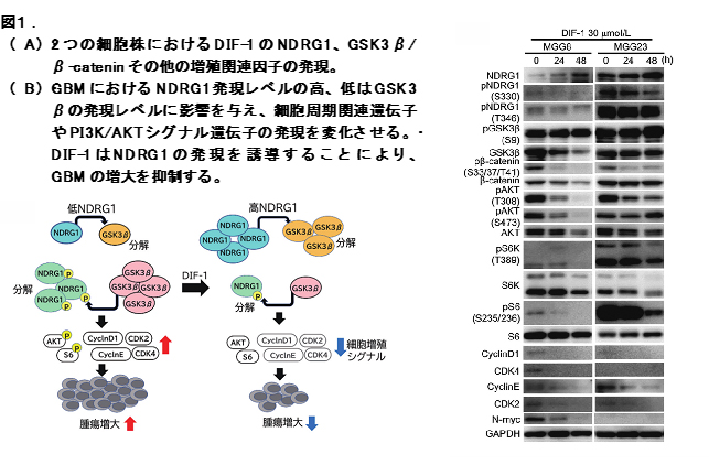
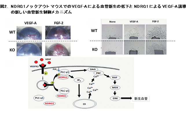
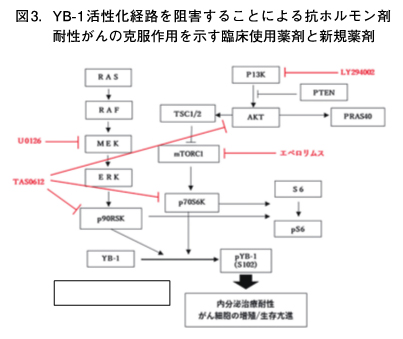
- Development of novel mechanisms and related biomarkers that are involved in progression and drug resistance in cancer
-
Elucidation of novel mechanisms and relevant biomarkers involving malignant progression and drug resistance in cancer can contribute to further improvement of anticancer therapeutics. Our present study uses three independent approaches to target N-Myc Downstream Regulated Gene 1 (NDRG1) and Y-box binding protein-1 (YB-1). First, enhanced expression of NDRG1 suppresses cell proliferation of glioblastoma (GBM) cells through inactivation of the GSK3β/β-catenin signaling pathway. Treatment with Differentiation Inducing Factor-1 (DIF-1) enhances NDRG1 expression, followed by suppression of cell proliferation and tumor growth by GBM in vitro as well as in vivo. Secondly VEGF-induced angiogenesis is specifically blocked in mouse cornea and aorta sprouting models for angiogenesis when NDRG1 is silenced. NDRG1 plays its critical role in VEGF-induced angiogenesis through activation of PLCγ1/ERK signaling pathway after VEGF binding to VEGFR in endothelial cells. Third, enhanced expression of YB-1 induces suppression of ERα expression in breast cancer cells, followed by the appearance of tumors resistant to antiestrogen therapeutic drugs. Treatment with inhibitors of PI3K/AKT and RAS/MEK/ERK pathways blocks YB-1 activation and overcomes resistance to antiestrogens in breast cancer.